")
Spinal specialists diagnose and treat various ailments of the spine. The medical profession tends to break the body down into parts so that doctors can specialize and understand everything there is to know about one area of the body. This can be helpful in diagnosing and treating a “symptom,†but ultimately, we will need to address the relationship between “all†the parts of the body if we aim to resolve the root cause of pain and dysfunction.
Many of my clients have seen specialists that diagnose a condition they believe is causing their pain. They are told they have excessive wear and tear, bulging discs, osteoarthritis, or compression. Unless there was an acute injury, my question is, “Why?†When the body is in alignment, there is less friction and compression that cause these symptoms; therefore, degeneration is less likely to occur. Muscles move bones, and in many cases, we can make muscular adaptations to take pressure off a joint or body part that’s compressed, dysfunctional, or out of alignment.
Back pain is the main symptom I hear complaints about, so I believe educating our selves on the structure of the spine and its various symptoms can be helpful to understand what’s causing them. In the article below, you will find more than you’ll ever need to know from a medical perspective about your spine and spinal symptoms.
The Spine
The spine is designed to protect your spinal cord. The spinal cord is a column of nerves that connect your brain with the rest of your body and control your movements. When the spinal cord is injured this exchange of information can be disrupted.
Your spine is made up of 24 small bones (vertebrae) that are stacked on top of each other to create the spinal column. Between each vertebra is a soft, gel-like cushion called a disc that helps absorb pressure and keeps the bones from rubbing against each other. Each vertebra is held to the others by groups of ligaments. There are also tendons that fasten muscles to the vertebrae. The spinal column has joints (just like the knee or elbow or any other joint) called facet joints. The facet joints link the vertebrae together and give them the flexibility to move against each other.
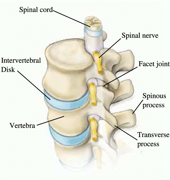
Each vertebra has a hole in the center, so when they stack on top of each other they form a hollow tube that holds and protects the spinal cord and its nerve roots. The spine branches off into thirty-one pairs of nerve roots. These roots exit the spine on both sides through spaces between each vertebra called neural foramina.
Spinal Segments and Curves
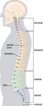
The spine itself is made up of the cervical spine, the thoracic spine, and the lumbar spine. The cervical is the upper part of the spine, made up of seven vertebrae. The thoracic is the center portion of the spine, consisting of 12 vertebrae. The lower portion of the spine is called the lumbar spine. It is usually made up of five vertebrae, however, some people may have six lumbar vertebrae. Below the lumbar spine is the sacrum. The sacrum is a group of specialized vertebrae that connect the spine to the pelvis.
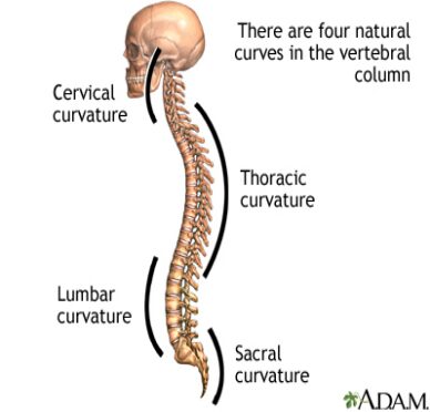
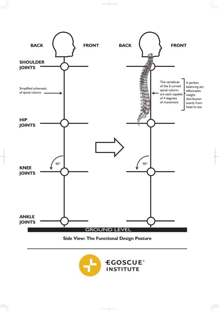
A functional spine has an “S” shaped curve when looking at it from the side. This allows for an even distribution of weight. The “S” curve helps a healthy spine withstand all kinds of stress. The cervical spine curves slightly inward, the thoracic curves outward, and the lumbar curves inward. Even though the lower portion of your spine holds most of the body’s weight, each segment relies upon the strength of the others to function properly.
This concept is important to recognize because injections and surgery do not influence spinal curves. The lumbar spine, for instance, is designed to be in a slight amount of extension. If there is excessive flexion or excessive extension (often due to the position of the hips and pelvis), back pain and compressive disc issues are more likely. Therefore, addressing the tilt of the pelvis may be necessary to reduce compressive forces in the spine and prevent further issues.
Vertebrae
The individual bones of the spine are called vertebrae. These are the building blocks of the spinal column. The vertebrae protect and support the spinal cord. They also bear most of the weight put upon your spine. The body of each vertebra is the large, round portion of bone and is attached to a bony ring. When the vertebrae are stacked on top of each other, this ring creates a hollow tube where the spinal cord passes through.
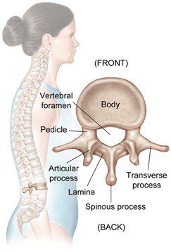
The bony ring attached to the vertebral body consists of several parts. First, the laminae extend from the body to cover the spinal canal, which is the hole in the center of the vertebrae. Second, the spinous process is the bony portion opposite the body of the vertebra. You feel this part if you run your hand down a person’s back. Then there are two transverse processes (little bony bumps), where the back muscles attach to the vertebrae. Finally, the pedicle is a bony projection that connects to both sides of the lamina.
Intervertebral Disc
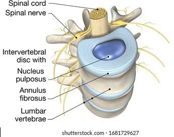
The intervertebral discs are flat, round “cushions” that act as shock absorbers between each vertebra. Each disc has a strong outer ring of fibers called the annulus, and a soft, jelly-like center called the nucleus pulposus. The annulus is the disc’s outer layer and the strongest area of the disc. The annulus is a strong ligament that connect the vertebra together and helps keep the disc’s center intact. The nucleus is made up of tissue that is very moist because it has high water content. The water content helps the disc act like a shock absorber.
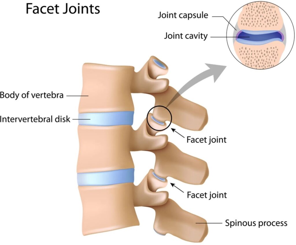
Facet Joints
The facets are the “bony knobs” that meet between each vertebra to form the facet joints that join your vertebrae together. There are two facet joints between each pair of vertebrae, one on each side. They extend and overlap each other to form a joint between the neighboring vertebra facet joints. Without the facet joints, you would not have flexibility in your spine, and you could only move in very straight and stiff motions.
The facet joints are what are known as synovial joints. A synovial joint, such as the knee or elbow, is a structure that allows movement between two bones. In a synovial joint, the ends of the bones are covered with a material called articular cartilage. This material is a slick, spongy material that allows the bones to glide against one another without much friction.
Surrounding the facet joint is a watertight sack made of soft tissue and ligaments. This sack creates what is called the “joint capsule”. The ligaments are soft tissue structures that hold the two sides of the facet joint together. The ligaments around the facet joint combine with the synovium to form the joint capsule that is filled with fluid (synovial fluid). This fluid lubricates the joint to decrease the friction, just like oil lubricates the moving parts of a machine.
Neural Foramen
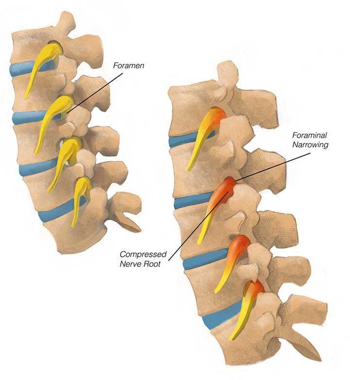
The neural foramen is the opening between every two vertebrae where the nerve roots exit the spine. The nerve roots travel through the foramen to reach the rest of your body. There are two neural foramina between each pair of vertebrae – one on each side. Without the foramen, nerve signals could not travel to and from the brain to the rest of your body and your body would be unable to function.
Spinal Cord and Nerve Roots
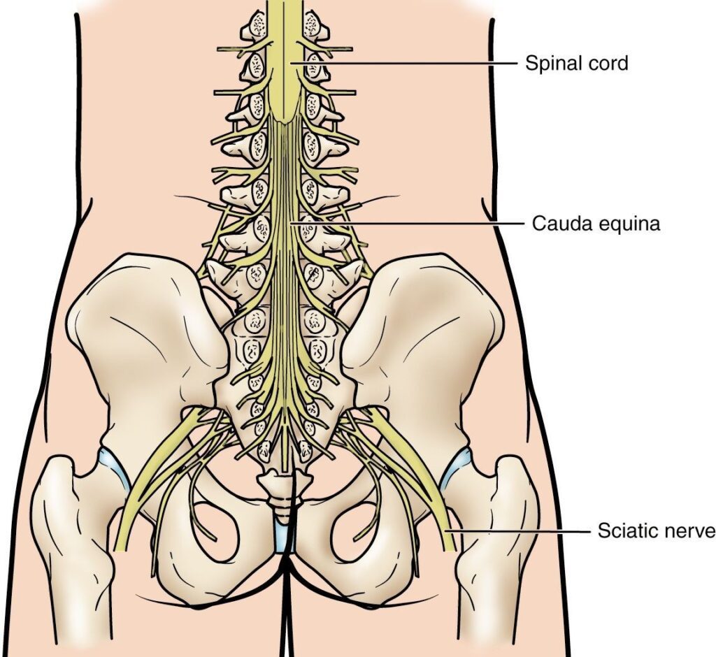
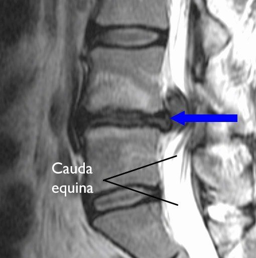
The spinal cord extends from the brain to the area between the end of your first lumbar vertebra and the top of your second lumbar vertebra. At the second lumbar vertebra, the spinal cord divides into several different groups of fibers that form the nerves that will go to the lower half of the body. For a small distance, the nerves travel through the spinal canal before exiting out the neural foramen. This collection of nerves is called the cauda equina while it is still inside the spinal canal.
A protective membrane called the dura mater covers the spinal cord. The dura mater forms a watertight sack around the spinal cord and the spinal nerves. Inside this sack, the spinal cord is surrounded by spinal fluid.
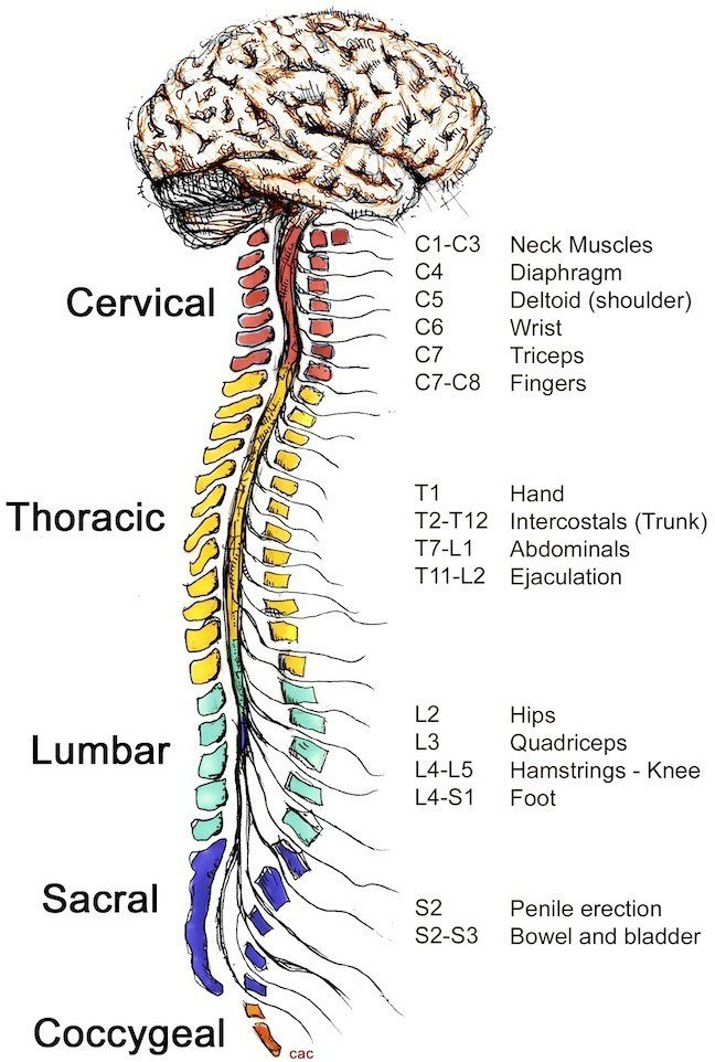
As previously mentioned, the nerve fibers in your spinal cord branch and travel through the foramina between your vertebrae. The nerves in each area of the spinal cord connect to specific parts of your body as seen in the illustration below. Consequently, damage to the spinal cord can cause paralysis in certain areas depending on which spinal nerves are affected. The nerves of the cervical spine go to the upper chest and arms. The nerves in your thoracic spine go to your chest and abdomen. The nerves of the lumbar spine then reach to your legs, bowel, and bladder. These nerves coordinate and control all the body’s organs and parts, and let you control your muscles.
Your nerves carry electrical signals back to the brain that enable you to feel sensations. Damage to the nerves themselves can cause pain, tingling, or numbness in the area where the nerve travels. When narrowing occurs in the spinal canal it can cause compression on the spinal cord and nerve roots. This is called stenosis and can result in numbness often in the hands and feet.
STENOSIS
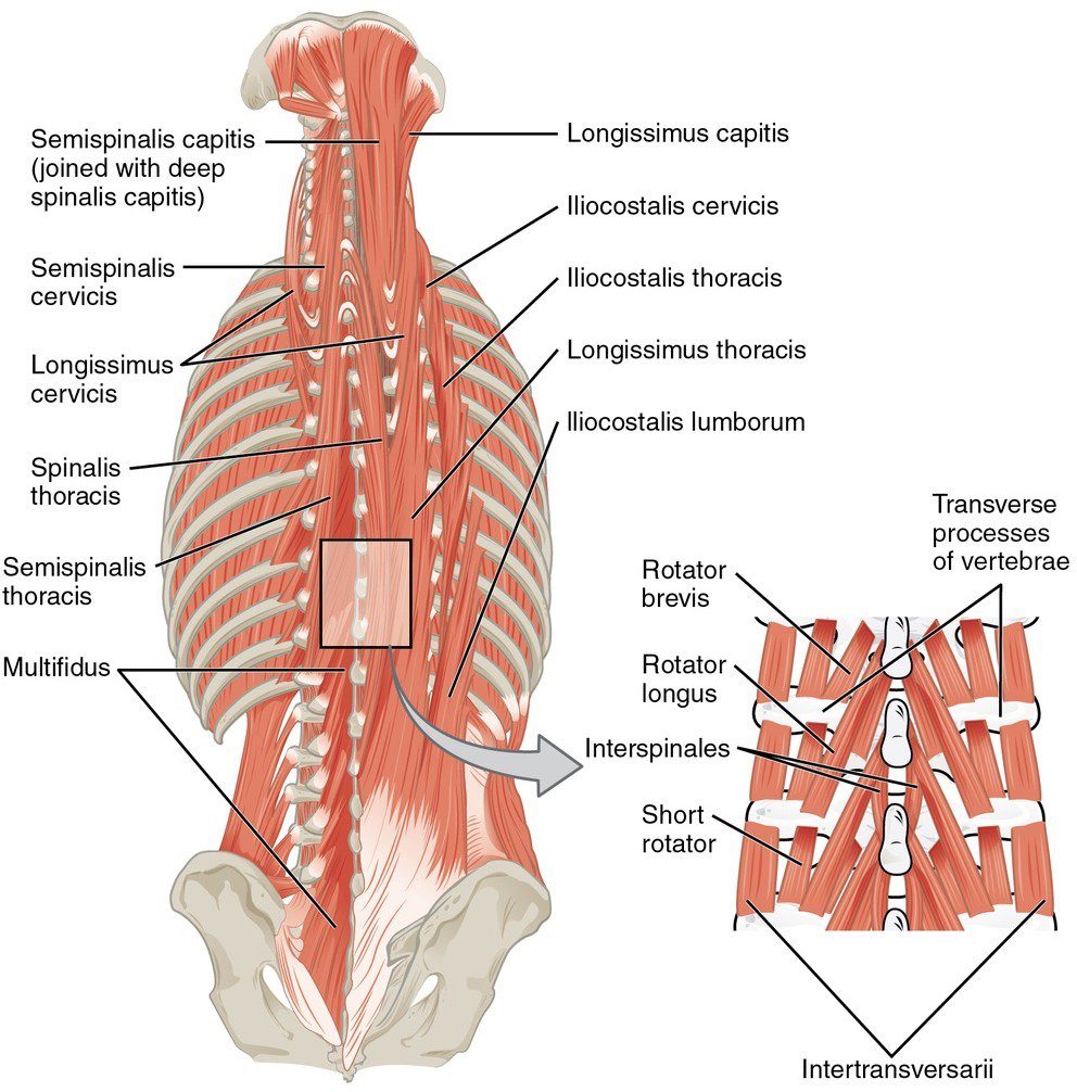
Paraspinal Muscles
The paraspinal muscles refer to the muscles next to the spine. They support the spine and the movement of the spine. Your joints allow for flexibility and your muscles allow for mobility. There are many small muscles in the back, each controlling some part of the movement between all the vertebrae and the rest of the skeleton.
These muscles can be injured directly, such as when you have a pulled muscle or muscle strain. The muscles can also cause problems indirectly, such as when the muscles are in spasm often due to an injury in another part of the spine.
Muscle Spasms
When you experience a muscle spasm, it is because your muscle tightens up and will not relax. These spasms usually occur as a reflex, meaning that you cannot control the contraction of the muscles. When any part of the spine is out of alignment or injured, the muscles may go into spasm to reduce motion around the area. This is a protective mechanism designed to protect the body.
When muscles are in spasm, they produce too much of the chemical, lactic acid. Lactic acid is a waste product produced by the chemical reaction inside muscle cells that allow the muscle to contract. If the muscle cell cannot relax, too much lactic acid builds up inside the muscles. The buildup of lactic acid causes a painful burning sensation.
Spinal Segment
A spinal segment is made up of the two vertebrae attached together by ligaments, with the disc separating them. The facet joints fit between the two vertebrae, allowing for movement, and the neural foramen between the vertebrae allow space for the nerve roots to travel freely from the spinal cord to the body.
Each spinal segment is like a well-tuned part of a machine. All the parts should work together to allow weight bearing, movement, and support. When all the parts are functioning properly, the spinal segments join to make up a remarkably strong spinal column. When one segment deteriorates to the point of instability, it can lead to problems throughout the entire spine causing pain and other difficulties. What’s important to recognize, is each of these parts/segments is influenced by the position of all the other bones in the body.
Now that we are familiar with the parts of the spine, let’s look more closely at the three segments of the spine: Lumbar, thoracic, and cervical:
Lumbar Spine
The lowest part of the spine is called the lumbar spine. This area has five vertebrae. However, sometimes people are born with a sixth vertebra in the lumbar region. The base of your spine (sacrum) is a fusion of many bones, and when one of them forms as a vertebra rather than part of the sacrum, it is called a transitional (or sixth) vertebra. This occurrence is not dangerous and does not appear to have any serious side effects.
The lumbar spine’s shape has what is called a lordotic curve. The lordotic shape is like a backwards “C”. When a part of the spine is in this shape it’s termed as being in extension or lordodic. When it’s shaped like a forward C it’s termed as being in flexion or kyphotic. If you think of the spine as having an “S”-like shape, the lumbar region would be the bottom of the “S”.
The vertebrae in the lumbar spine area are the largest of the spine, so the lumbar spinal canal is larger than in the cervical or thoracic. Because of its size, the lumbar spine has more space for the nerves to move about. The larger the vertebrae, the more stable that area of the spine is designed to be.
Your pelvis is the core of your body and where most healthy movement is generated. The hips and pelvis make up your center of gravity. Lower back pain is likely one of the most common complaints due to the fact the lumbar spine is directly connected to your pelvis. Although most movement should be occurring at the hips and pelvis, if there is restriction, excessive instability or immobility, more pressure may be put on the lumbar spine. As a result, injuries can be common when lifting, twisting, or carrying heavy loads.
Thoracic Spine
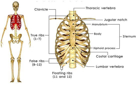
The thoracic spine is made up of the middle 12 vertebra of the spine. These vertebrae connect to your ribs and form part of the back wall of the thorax (the ribcage area between the neck and the diaphragm).
This part of the spine has very narrow, thin intervertebral discs, so there is much less movement allowed between vertebrae than in the lumbar or cervical parts of the spine. It also has less space in the spinal canal for the nerves. The thoracic spine’s curve is called kyphotic because of its shape, which is a regular “C”-shaped curve with the opening of the “C” in the front.
Cervical Spine
The cervical spine is made up of the first seven vertebrae. It starts just below the skull and ends just above the thoracic spine. The cervical spine has a lordotic curve (a backward “C”-shape) – just like the lumbar spine. The cervical spine is much more mobile than the other spinal regions – think about all the directions and angles a functional neck turns.
Unlike the rest of the spine, there are special openings in each vertebra in the cervical spine for the arteries (blood vessels that carry blood away from the heart), as well as the spinal canal that carries the spinal cord. The arteries that run through these openings bring blood to the brain.
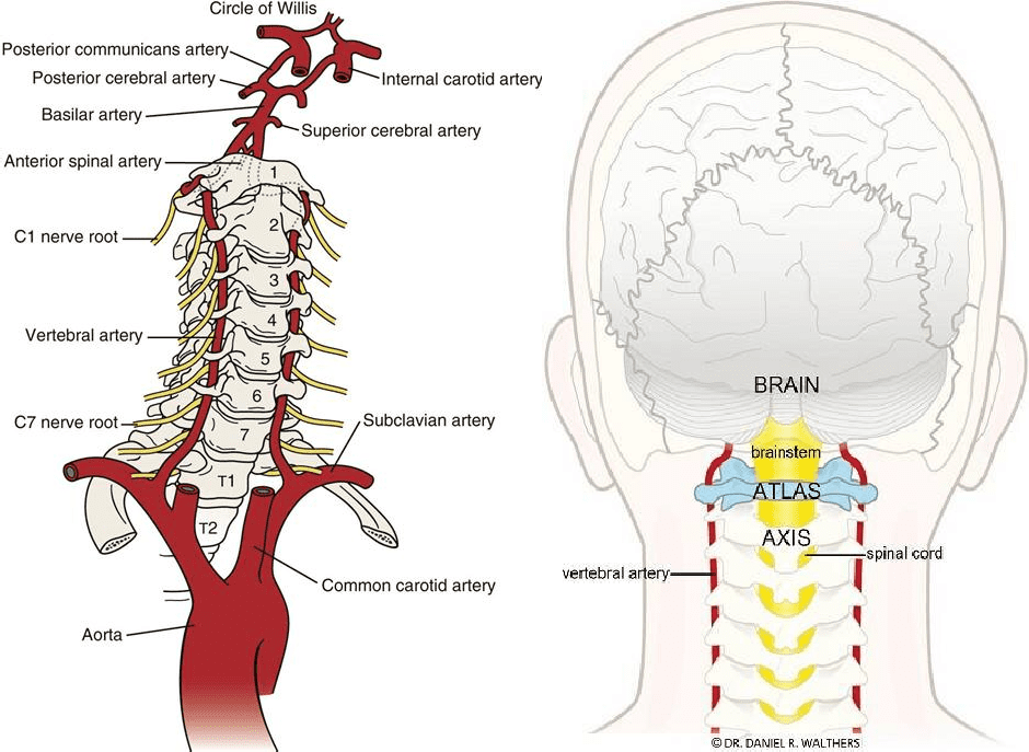
Two of the vertebrae in the cervical spine: the atlas and the axis, differ from the other vertebrae because they are designed specifically for rotation. These two vertebrae are what allow your neck to rotate in so many directions, including looking to the side.
The atlas is the first cervical vertebra – the one that sits between the skull and the rest of spine. The atlas does not have a vertebral body but has a thick forward (anterior) arch and a thin back (posterior) arch, with two prominent sideways masses.
The atlas sits on top of the second cervical vertebra – the axis. The axis has a bony knob called the odontoid process that sticks up through the hole in the atlas. It is this special arrangement that allows the head to turn from side to side as far as it can. Special ligaments between these two vertebrae allow a great deal of rotation to occur between the two bones.
Though the cervical spine is very flexible, it is also very much at risk for injury from strong, sudden movements, such as whiplash type injuries. This high risk of harm is due to the limited muscle support that exists in the cervical area, and because this part of the spine supports the weight of the head. This is a lot of weight for a small, thin set of bones and soft tissues to bear. Therefore, sudden, strong head movement can create injury to the cervical spine.
Mechanical & Compressive Pain
It is useful to understand back issues by dividing them into two different categories: Mechanical and compressive back pain.
Mechanical pain is linked with the movement, or “the mechanics” of the spine. This type of pain occurs when injury to the spine’s discs, facet joints, ligaments, or muscles results in inflammation. When there are faulty mechanics, the more you use the back, the more it hurts. Compensatory biomechanics can lead to conditions such as fractures of the vertebra, muscle strains, ligament injures in the spine, and wear and tear of the spine’s joints and discs,
On the other hand, compressive pain is a result of pressure or irritation on the spinal cord, or nerves that leave the spine. For example, if an intervertebral disc herniates (usually called a ruptured disc) and pushes into the spinal canal, it can cause compression on the nerve. This pressure or irritation can cause pain, numbness, and muscle weakness where the nerve travels.
Although it can be helpful to understand whether the pain is coming from a compressive or mechanical problem, it can also be helpful to shift the focus away from the spine and onto the hips or shoulders. Dysfunction in either of these areas will influence the friction, mobility, and stability of each of the spinal segments.
In the next segment of this article, we will discuss various spinal symptoms and diagnoses resulting from mechanical and compressive dysfunction:
MEDICAL DIAGNOSES
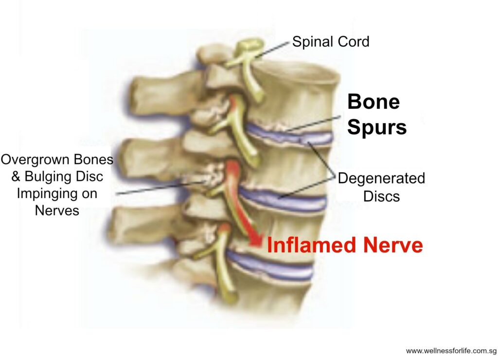
Bone Spurs
The body may grow bone to protect the spine when there is excessive friction. This is called a bone spur and can impinge upon the nerves leading to pain and irritation. Bone spurs grow as a protective mechanism, so unless you determine what’s causing friction along the joints of the spine, the body will most likely continue to find a way to protect itself, even if you surgically remove the symptomatic part.
Arthritis & Osteoarthritis
The term arthritis means inflammation of the joints. Arthritis of the spine usually refers to a condition where there is inflammation of the facet joints between the vertebrae. It’s important to recognize there are two types of arthritis: Systemic inflammatory arthritis, and wear- and-tear arthritis. A systemic type of arthritis is a disease process that affects all the joints of the body. Rheumatoid arthritis is one such disease. The joints of the spine may be involved in systemic types of arthritis because the facet joints are made up of the same tissues as any other joint. Therefore, diseases that attack joint tissues also attack the facet joints.
Wear-and-tear arthritis, also called osteoarthritis, can come from a single injury that damages the joint. or it can result from a lifetime of overuse of different joints that damage the joint a little bit at a time.
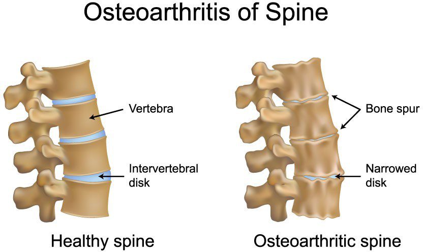
Osteoarthritis is caused by a breakdown of the articular cartilage inside the affected joint. Articular cartilage is the material inside the joint that cushions the bones of the joints from impact and allows smooth, gliding motions. Damaged cartilage begins to fray, making it less flexible and more prone to injury. Over time, the cartilage can wear away completely, causing the bony surfaces of the joint to rub directly against each other. Eventually the joint becomes worn away and bone spurs develop around the joint to protect it.
Facet Joint Syndrome
In many cases, dysfunction of the facet joints can contribute to back pain. When your doctor thinks the facet joints are a major source of your pain, he may use the term “facet joint syndrome”. Facet joint degeneration, or osteoarthritis, can be caused by a combination of aging, pressure overload of the facet joints, faulty biomechanics, or previous injury.
When there is degeneration of the discs, this can create pressure overload on the facet joints. As the discs degenerate, they wear down and begin to collapse. This narrows the space between each vertebra. This narrowing of the space between each vertebra affects the way the facet joints line up. When this occurs, it places too much pressure on the articular cartilage surface of the facet joint. The excessive pressure leads to damage of the articular surface and eventually the cartilage begins to wear away.
When facet joint arthritis gets bad enough, the cartilage and fluid that lubricate the facet joints are eventually destroyed, leaving bone rubbing on bone and bone spurs begin to form around the facet joints to protect the spine.
When bone spurs develop, they can take up space in the foramen (the opening between vertebrae where nerve roots exit the spine) and press into nerve roots. As the bone spurs begin to grow larger, they can eventually extend into the spinal canal itself. This leads to narrowing of the spinal canal called spinal stenosis.
Radiculopathy
Radiculopathy is the medical term used to describe a “pinched nerve” in the spine. Radiculopathy occurs when a nerve is irritated by something that is either rubbing on the nerve or pressing on the nerve. In some cases, such as a herniated (or ruptured) disc, there may also be a chemical reaction irritating the nerve. Chemicals released from the inside of the disc seem to irritate nerve tissue, causing pain and inflammation of the nerve.
Abnormal pressure or irritation on a particular nerve causes several problems. First, there is numbness in the area where the nerve is designed to provide sensation or feeling. For example, if the nerve ends in the side of the foot and supplies sensation to that area, it will have decreased feeling or pain.
The key to understanding radiculopathy is understanding that while the irritation or pressure on the nerve may be in your back, your brain thinks the pain is coming from another part of your body such as your foot or hand. You may have pain or weakness in the muscles; furthermore, the reflexes controlled by the muscles may not work. By determining which reflexes are not working, a doctor can usually tell which nerve is involved with the problem. Radiculopathy can be caused by herniated discs, bone spurs, tumors, and fractures that put pressure on the nerves.
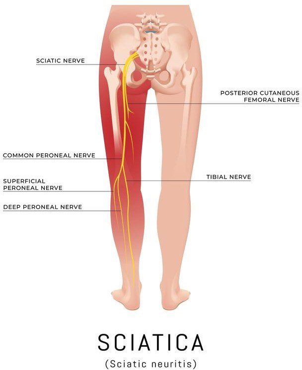
Sciatica
The term sciatica refers to a certain type of radiculopathy that occurs in the leg. It is called sciatica because it describes the radiculopathy that occurs when one or more of the nerves that make up the large sciatic nerve are irritated or pinched. Sciatica is not any different than a pinched nerve anywhere else in the spine, it simply has its own name because it is more common than other nerve conditions. It occurs in the lumbar spine, which tends to be the most common site of spinal nerve irritation.
Sciatica describes the pain that travels from the sciatic nerve in the lumbar region into your buttocks, back of the thighs, and sometimes calf and foot. The pain is typically caused by irritation of the nerve roots that join outside the spine to make up the sciatic nerve. Some conditions that can cause sciatica are bulging or herniated discs, bone spurs, cancerous tumors that are growing into the nerves, and fractures that put pressure on the nerves.
To understand sciatica more clearly, let’s look at how the body works form the foot up. The muscle that’s responsible for flexing the foot is the tibialis anterior (the front shin muscle). The peroneal nerve connects to the tibialis anterior up to the knee where it branches into the sciatic nerve. The sciatic nerve travels through the pelvis and into the buttocks where the five nerve roots group together near the piriformis muscle. The sciatic nerve then connects to the sacral vertebrae S1, S2 and S3. The sacrum (the base of the spine) connects to the illum (the pelvic bone) constituting the SI joint where these nerves cross over. The sciatic nerve then connects to the vertebrae of the lower back at L5 and L4. This helps to clarify how a bulging lumbar disc can cause pain, tingling or numbness in the calf or foot. It also is indicative as to why it’s imperative in many cases to realign the position of the pelvis in order to receive sciatic pain.
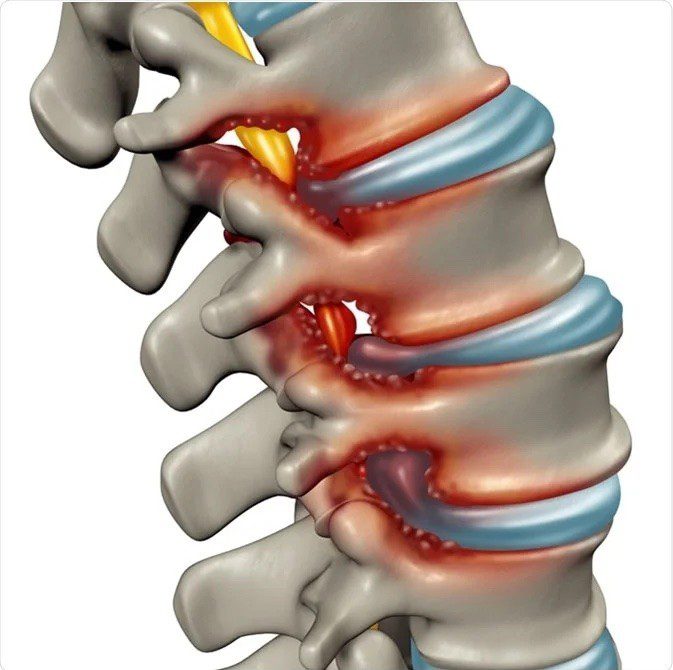
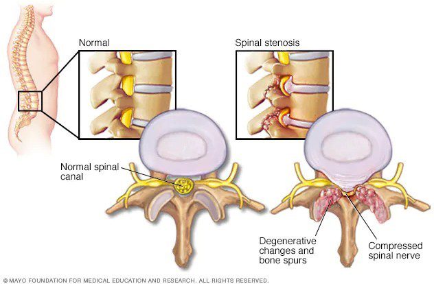
Spinal Stenosis
We have seen how individual nerve roots are affected by pressure and irritation — but what about pressure on the spinal cord itself?
Pressure on the spinal cord typically results from stenosis. The definition of stenosis is the narrowing of an opening or tube – in this case being the spinal canal. Stenosis can occur in all areas of the spine, but it is most common in the cervical and lumbar spine.
Bone and tough ligaments surround the spinal canal. If anything begins to narrow the spinal canal, the risk of irritation and injury of the spinal cord or nerves increases. Some conditions that can lead to narrowing of the spinal canal include infection, tumors, trauma, a herniated disc, arthritis, thickening of ligaments, growth of bone spurs, and disc degeneration.
Spinal stenosis usually occurs in older people after years of wear and tear or degeneration of the spine stemming from dysfunctional biomechanics. This wear and tear results in changes in the structures around the spinal canal, such as thickening of the large ligaments that connect the vertebra together, bone spurs around the facet joints, and bulging of the discs themselves. This causes a push into the spinal canal, making the tube of the spinal canal smaller.
Eventually, there is not enough space in the spinal canal for the nerve to comfortably fit without causing too much pressure. Stenosis can also develop because of injuries, infections, or tumors. Some people even have a narrow spinal canal from birth.
This lack of space also causes the supply of blood and oxygen to the spinal cord to be reduced. When the spine needs more blood flow during increased activity, the blood vessels may not be able to swell to get more blood to the spine. This can lead to numbness and pain in the affected nerves. The nerves also lose some of their mobility when they don’t have enough space, which can lead to irritation and inflammation.
Other symptoms of spinal stenosis include a sensation of heaviness, weakness, and pain when walking or standing for long periods. With rest, these symptoms often disappear. These symptoms occur because the nerve roots are being aggravated, upsetting the normal signals that travel from the brain to the body. Irritation of the nerves in the spinal canal is often worse when standing or walking because of the mechanical compression and stretching of the nerves, whereas in some cases, sciatica is often worse when sitting due to the increased compression on the nerve.
Discogenic Pain
Discogenic pain is a term back specialists use when referring to pain caused by a damaged intervertebral disc. With a degenerative or bulging disc, movements that place stress on the disc may result in back pain. There is some evidence that this may come from the disc itself. Just as with any other body part that is injured, such as a broken bone, or even a cut in the skin, when these types of injuries are immobilized, there is a reduction in pain.
Discogenic pain usually causes pain felt in the lower back. It may also feel like the pain is coming from your buttock areas and even down into the upper thighs. The experience of feeling pain in an area away from the real spot causing the pain is common in many areas of the body, not just the spine. Examples include: a person who has gallstones may feel the pain in their shoulder; or a person experiencing a heart attack may feel pain in the left arm. This is called radiating pain. It is very common for pain produced by spinal problems to be felt in different areas of the body other than the spine itself.
Bulging Discs
Bulging discs are relatively common and are not a cause for panic. In fact, abnormalities that show up on MRIs, such as bulging or protruding discs, are often seen in patients both with and without back pain. Most likely, discs begin to bulge due to degenerative processes resulting from bony misalignments and faulty biomechanics.
A bulging disc becomes painful when it bulges enough to cause a narrowing of the spinal canal. If there are bone spurs present on the facet joints behind the bulging disc, the combination may cause narrowing in that area leading to segmental spinal stenosis.
Degeneration of the Intervertebral Disc
The process of disc degeneration can cause many problems in the spine. When the load bearing joints lose their proper alignment, just standing upright will test the spine’s ability to support your body weight. Over time, these repetitive stressors add up and begin to affect the discs in your spine. Minor injuries to a disc may occur and not cause pain at the time; however, as they add up, the disc eventually begins to suffer from the wear and tear and begins to degenerate.
The intervertebral discs are designed to absorb pressure and keep the spine flexible by acting as cushions during body movement. The discs work similarly to shock absorbers. They are like cushions in running shoes; without them, a jogger would feel every pound on the pavement, and the feet would soon tire out. Without the cushioning effect of the discs, the vertebrae in your spine would probably fracture or break. Bones cannot sustain high stress repeatedly without being cushioned.
Disc space and bone spurs
A healthy intervertebral disc has a great deal of water in the nucleus pulposus – the center portion of the disc. The water content gives the nucleus a spongy quality and allows it to absorb spinal stress. Excessive pressure or injuries to the disc can cause the injury to the annulus – the outer ring of tough ligament material that holds the vertebrae together. Small tears show up as in the ligament material of the annulus. These tears heal by developing scar tissue; however, the scar tissue is not as strong as normal ligament tissue. Over time as more scar tissue forms, the annulus becomes weaker. Eventually this can lead to damage of the nucleus pulposus. The nucleus begins to lose its water content and it begins to dry up.
Because of water loss, the discs lose some of their ability to act as a cushion. This can lead to even more stress on the annulus and still more tears as the cycle repeats itself. As the nucleus loses its water content, it collapses allowing the two vertebrae above and below to move closer to one another. This results in a narrowing of the disc space between the two vertebrae. As this shift occurs, the facet joints located at the back of the spine are forced to shift. This shift changes the way the facet joints work together.
The body attempts to avoid the excess motion and friction at the dysfunctional spinal segment by developing bone spurs, also called osteophytes. These bones spurs can also form around the facet joints. The bone spurs become a problem when they begin to grow into the spinal canal and cause spinal stenosis irritating the nerves.
What is important to recognize is many of these symptoms are the body’s protective mechanism. When we surgically remove the bone spur, we are not addressing what is causing the friction or excessive motion at that spinal segment.
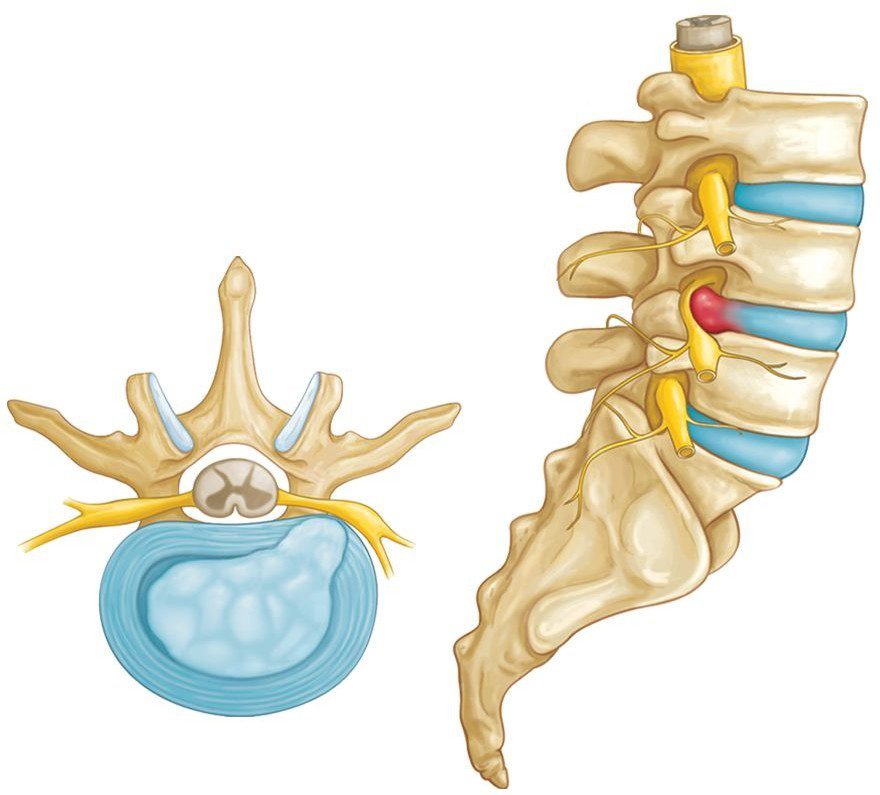
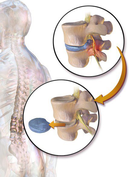
Herniated Disc
A herniated disc occurs when the intervertebral disc’s outer fibers (the annulus) are damaged and the soft inner material of the nucleus pulposus ruptures out of its normal space. If the annulus tears near the spinal canal, the nucleus pulposus material can push into the spinal canal. This can cause excessive pressure on the spinal cord and nerve roots. There is also some evidence that the nucleus pulposus material causes a chemical irritation. Both the pressure on the nerve root and the chemical irritation can lead to problems with how the nerve root works. The combination of the two can cause pain, weakness, and/or numbness in the area to which the nerve travels. For this reason, a herniated disc usually causes pain of the compressive type. Sometimes a herniated disc is referred to as “slipped disc”, though the disc does not actually slip.
Herniated discs are most common in the lumbar spine because of all the pressure it supports being closest to the center of the body. A herniated lumbar disc will often produce sciatic symptoms felt as pain in the lower back and numbness radiating down the back of the leg, side of the calf, and possibly into the side of the foot. In some cases, you may not necessarily have much back pain. The exact area where you will feel numbness depends on the nerve root that is affected; the numbness could be in the inner ankle, big toe, heel, outer ankle, outer leg, or a combination. Medical professionals use this information to get an idea which nerve is affected.
Pressure on the nerve root can cause the nerve that controls the muscles not to work properly. This can result in weakness of some muscles and may change the reflexes in certain areas. For example, a disc compression in the neck may result in pain that is perceived in the arm or hand.
Herniated Disc
A herniated disc is much less common in the thoracic spine. This is likely because the discs are much thinner and there is less material in the nucleus pulposus to rupture into the spinal canal. However, if a herniated disc does occur in the thoracic spine, it can be much more serious than in the lumbar spine. The thoracic spine has very little extra room in the spinal canal. In addition, a herniated disc in the thoracic spine puts pressure on the spinal cord – not just a few nerve roots. Too much pressure on the spinal cord from a herniated thoracic disc can lead to total paralysis from the waist down.
In most cases, a herniated disc will not need surgery. The treatment of a herniated disc depends on the symptoms. It also depends on whether the symptoms are getting steadily worse — or whether they are getting better. In many cases, the initial problems due to a herniated disc resolve over several weeks to months with no intervention. To speed up the process finding positions that take pressure off the nerve and avoiding positions that compress the nerve can speed up recovery.
I often compare this healing to a broken leg. Healing requires patience and adequate time. Healing does not typically happen overnight but symptoms should get adequately better over time. If you reduce any motion that irritates the symptom for a short period of time as it heals and then slowly reintroduce proper movement, the body has a natural capability to heal on its own without intervention. In cases where it worsens over time, it’s best to consult a medical professional.
Cauda Equina Syndrome
In rare cases, a herniated disc in the lumbar spine area can be so large that it fills the entire spinal canal in the area where it ruptures. When the spinal canal fills with disc material, it may place a great amount of pressure on the nerves. If this occurs in the lower spine, it can lead to a condition called “cauda equina syndrome”. This problem can lead to permanent paralysis of the muscles that control your bowels and bladder. If you lose control over your bladder or bowels, this can be quite serious and often requires medical attention.
Spondylolysis and Spondylolysthesis
A spondylolysthesis is a condition where one vertebra shifts forward of another and usually occurs between the L5 and S1 vertebrae. It’s often associated with spondyloysis which is a fracture of the pars articularis, which is a portion of the vertebral arch as seen below.
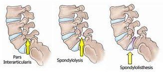
Pars Articularis Fracture
The fracture of the Pars Articularis, otherwise known as spondylolysis, causes instability in the lumbar (lower back) region and often is associated with pain, especially upon pelvic/lumbar extension (arching of the lower back). Spinal alignment is dependent upon both the disc and the facet joints to anchor the vertebrae back. When there is a fracture, the facet joints can’t prevent the vertebrae from being pushed forward and the disc slowly stretches and allows the upper vertebrae to slide forward. This often results in lower back pain and can also lead to numbness in the glutes or referred pain or numbness in the leg or foot. With spondylolisthesis surgery is rarely an option. More often, corrective exercise and physical therapy are prescribed to alleviate the symptoms.
Segmental Instability of the Spine
Each spinal segment is like a well-tuned part of a machine. The parts should work together to allow weight bearing, movement, and support. When all the parts are functioning properly, the spinal segments join to make up a remarkably strong spine. However, when one segment deteriorates to the point of instability, it can lead to localized pain and difficulties.
Segmental instability occurs when there is too much movement and friction between two vertebrae. When the spine is unable to move as it’s designed, the vertebrae may be forced to compensate, picking up this task they are not designed for. Excess movement of the vertebrae can cause pinching or irritation of nerve roots and too much pressure on the facet joints, leading to inflammation. It also may cause muscle spasms as the paraspinal muscles try to stop the spinal segment from moving too much. This instability eventually results in faster degeneration of the spine.
As the disc continues to degenerate, the facet joints can become arthritic, bone spurs form around the joints, and the segmental instability gets worse. It’s imperative to put a stop to this cycle by restoring alignment as well as proper mobility in specific areas of the body designed to be mobile, so they areas that are meant to be stable can be restored to perform as designed.
MEDICAL TREATMENTS
If you visit a back specialist, their job is to diagnose and determine exactly how serious your symptom is. Some issues need immediate attention; however, most back problems will not require surgery.
A variety of medical treatment options exist for different types of back pain, and in most cases, simple therapies such as mild pain medications, rest, and time, are effective in relieving the immediate pain symptoms. The overall goal of most medical treatments is: To make you comfortable as quickly as possible, to design a program to reduce further degeneration, and to get you back to normal activity as soon as possible. The more you know about how your back works and what you can do to prevent further injury, the more effective your program will be. Below are descriptions of the most common forms of medical treatments, along with a brief explanation of what each is designed to do.
“Conservative” Treatment
Back specialists often use the term “conservative treatment” to describe any treatment option that does not involve surgery. Therefore, you may read that your provider is recommending a course of conservative treatment for your back problem. Treatment for your back pain may be as simple as reassuring you that it is not a serious problem and doing nothing but being patient while it heals.
However, usually anyone who has a back problem that becomes symptomatic should consider some preventive measures. Once adequate rest and healing has taken place, exercises to restore mobility, stability, and alignment should be considered. These exercises can be quick and easy to do, do not require any special equipment, and can help prevent future problems.
Rest and Recovery
As previously mentioned, immediately after a back injury, rest and patience is often all your back needs to feel better. Adequate rest allows for healing and takes the pressure off your spine and the muscles around your spine. Placing a pillow under your knees is often helpful to relieve pain and take pressure off the spine. However, do not stay in bed for several days! Bed rest for more than two or three days can weaken the back muscles, making the problem worse instead of better. Even though you may still feel some pain, a gradual return to normal activities is good for your muscles. In most cases of sudden back pain, the sooner you start moving safely again, the sooner your back pain will resolve. If you are sent to see a physical therapist, the first few days may be spent educating you on ways to take stress off the back while remaining as active as possible. Short periods of rest combined with brief exercises that do not increase your pain symptoms is the most helpful form of recovery.
Medication:
Doctor’s will often prescribe mild pain medications to reduce inflammation and pain. Medications will not address the cause of your back pain, but in cases of acute symptoms, they can help to control the pain while you begin to address what’s causing the symptom.
NSAIDs (Non-Steroidal Anti-Inflammatory Drugs)
Your doctor may prescribe NSAIDs or over-the-counter pain relievers like iibuprofen or naproxen. NSAIDs can be effective in relieving the pain associated with muscle strain and inflammation. They block the inflammatory response in joints. However, be aware that NSAIDs can decrease renal function and excessive use can lead to kidney problems. And once again, they do not address what is causing your symptoms and should only be used to reduce acute pain for short periods of time.
Epidural Steroid Injections (ESI), Cortisone injections and Nerve Blocks
An epidural can be used to relieve the pain of irritated nerve roots. The injection can also help reduce swelling from a herniated or bulging disc. The steroid injections are a combination of cortisone (a powerful anti- inflammatory steroid) and a local anesthetic that are given through an epidural into the spine. Epidural steroid injections are not always successful in relieving symptoms of inflammation. Medical professionals are trained to prescribe them only when conservative treatments have failed; and once again, they don’t address what’s causing the symptom and can be harmful when overused.
In clinical trails they found that although cortisone shots significantly reduced pain in some cases, over 50% of people experience a relapse of symptoms within 6 months of the injections. These studies also found that people who received cortisone injections had a lower rate of full recovery than people who did nothing at all. The alarming result of this study was people who had the cortisone injections had a 63% higher risk of relapse the those who opted for physical therapy or rest and recovery. Another study showed that an average of 4 injections resulted in a 57% worse outcome when compared to one injection.
Cortisone shots do seem to provide pain relief, as they effect the neural receptors; however they do not heal the structural damage which is the underlying cause of the pain and in fact, may prevent structural healing to occur.
Physical Therapy:
If physical therapy is recommended, your treatment plan could include one or more of the following: Alternating heat and ice, massage, ultrasounds, and electric stimulation, bracing ranging from a simple corset to a rigid plastic body jacket, flexibility and strength training through exercises, stretching, etc., pool therapy which unloads spinal pressure due to the decrease in gravity provided by the water, and learning how to stand, sit, and move more efficiently.
SPINAL SURGERIES
Discectomy
A discectomy is when a doctor removes the damaged part of a disc to relieve the pressure on the spinal nerve. I must interject that even if removing the disc matter resolves the symptom, it does not treat the root dysfunction. If you don’t treat the cause of the symptom, being bony misalignments, compensatory patterns, and improper biomechanics it’s common for pain to reoccur.
A discectomy has not been proven to be helpful in treating all cases of back or neck pain and most people find relief with more- conservative treatments such as time, rest, and physical therapy; however, a discectomy is suggested by doctors when conservative treatments have not been successful, or symptoms continue to worsen.
Discectomy
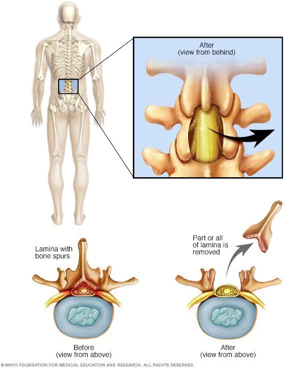
Laminectomy (decompression surgery)
A laminectomy is a surgery that creates space by removing the lamina. By removing the back part of the vertebra that covers the spinal canal the surgery is intended to relieve pressure on the spinal cord or nerves. This treatment is generally only used when more conservative treatments and rest have failed to relieve the symptoms. Most people see improvement in pain that radiates down the leg or arm, but less with pain in the back itself. A laminectomy is commonly used with spinal stenosis, as it removes the bone spurs that can be irritating the nerves of the spine. What we must recognize, is the friction that caused these bone spurs in the first place is not being addressed by performing this surgery. Unless the what’s causing this symptom is dealt with, it is likely to return at another spinal segment.
Laminectomy
Vertebroplasty and kyphoplasty
Vertebroplasty and kyphoplasty are similar procedures in which a hollow needle is passed through the skin of your back and into a fractured vertebra. In vertebroplasty a bone cement is injected into the fractured bone. In kyphoplasty, a balloon is first inserted and inflated to expand the compressed vertebra to its normal height before filling the space with bone cement. The hope is the cement strengthened vertebra will increase stability allowing the patient to stand straighter and reduce pain. Without treatment, fractures typically heal, but this surgery is designed to return the vertebra to its normal position before the bone hardens. However, studies show that people who get one fracture are five times more likely to develop additional fractures. This may be because the root dysfunction causing the fractures is not being addressed.
Foraminotomy
A foraminotomy is a surgery that widens the foraminal openings in your spine where the nerve roots exit the spinal canal. It’s typically recommended by doctors when there is a narrowing of the nerve openings (foraminal stenosis). It’s performed to potentially relieve symptoms of nerve root compression in cases where the foramen is being compressed by bone, disc, scar tissue or excessive ligament development that results in nerve compression. It’s designed to relieve pressure on the compressed nerves. An incision is made in the middle of the back of your spine. Skin, muscles, and ligaments are moved to the side and some bone is cut or shaved away to open the nerve root opening (foramen). From there, any disc fragments are removed. A full recovery can take 3-18 months.
Nucleoplasty (plasma disc decompression)
This is a treatment option for severe back pain resulting from a disc herniation that did not respond to conventional treatment. It’s an outpatient procedure that uses a needle to emit radio waves to shrink a disc bulge by dissolving excess tissue. It’s designed to relieve pressure inside the disc and on the nerves responsible for causing pain.
Spinal fusion (spondylodesis)
Spinal fusion is a surgery that should be a last resort when all else has failed, or when a serious spinal injury has occurred that must be addressed immediately. It’s a surgical technique in which two or more vertebrae are fused. This will prevent any movement of the spine in that area. We must keep in mind that the spine is designed to move. In almost all cases of spinal fusion other compensatory patterns are likely to occur when one spinal segment becomes immobile. In many cases this procedure may address an acute symptom, but can lead to further issues in the future, as other parts of the spine will be forced to compensate for the immobile segments.
ALTERNATIVE THERAPIES
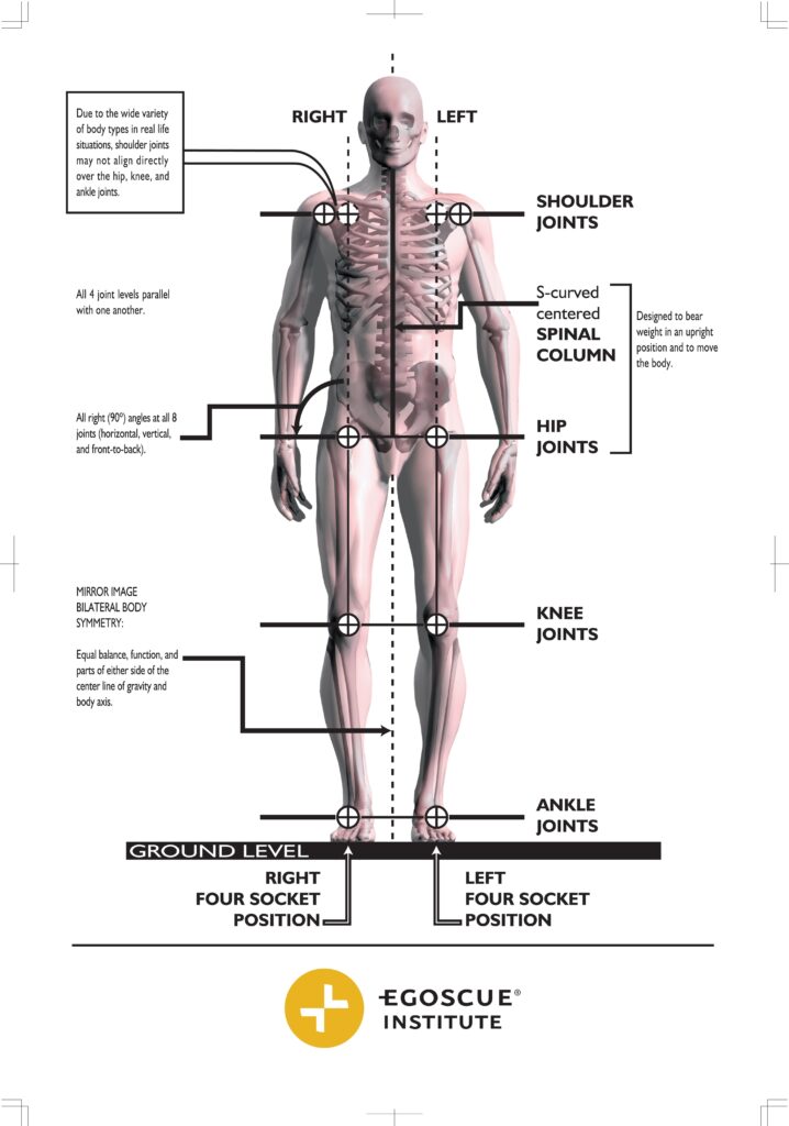
Although I recognize there are cases for the medical treatment protocols above, I take issue with one important aspect: Most are diagnosing and treating a “symptom†rather than addressing the underlying cause. There are a variety of alternative modalities that are trained to take a holistic view of the body. When you go to your foot, knee, hip, and spine doctor, this is often not the case. To treat the root dysfunction causing these symptoms, it’s often important to address the body as a whole unit. In the diagram below you can see how dysfunction of any joint might affect the ones above or below if it:
First metatarsal: mobile Mid foot: stable
Ankle: mobile
Knee: stable
Hip: mobile
Lumbar spine: stable Thoracic
spine: mobile Cervical spine
(lower): stable Cervical spine
(upper): mobile Shoulder: mobile
Elbow: stable
Wrist: mobile
Notice how stability and mobility alternate. If you want to improve knee stability, doesn’t it make sense you might also need to improve the mobility of the ankle and hip? Likewise, if you lose proper hip mobility, wouldn’t this impact the stability of the lumbar spine?
Below is an example of how this mobility/stability paradigm can influence the entire kinetic chain:
If one has lost mobility in the ankle, the foot may no longer flex and extend as it’s designed, and the knee is then forced to increase its mobility. This can lead to knee pain as the knee is designed to be a stable hinge joint and not excessively rotate. The hips in turn may lock up and the lower back, which should be stable, may become unstable due to the stiff hip joints. The thoracic spine and shoulder joints then engage to provide increased stability leading to potential upper back tightness and pain. The tightness and lack of mobility in the shoulder joints can also influence the flexors and extensors into the arm, which in turn can lead to symptoms such as carpal tunnel, tennis elbow or frozen shoulder. All these issues will alter the tilt of the pelvis and can create imbalances leading to rotations throughout the kinetic chain that ultimately cause friction and degeneration.
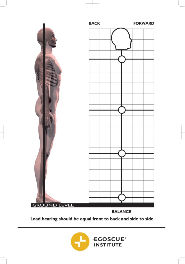
Back pain is often the result of faulty biomechanics that can stem from any joint or part of the body as reflected above. Addressing the vertical alignment of all these joints as well as imbalances between the right and left sides of the body, and restoring functional spinal curves, is often essential to ultimately fix what’s causing most pain symptoms.
Becoming educated, aware, and paying attention not only to your physical body, but your mental/emotional state, which has a strong influence over the nervous system is critical in alleviating these issues. Paying attention to what daily activities are exacerbating your pain such as prolonged sitting, repetitive stressors, improper exercise progressions, or mental/emotional stress that puts the body into a fight/flight mode is another important step in healing.
The recognition that each part of the body influences the whole and the whole influences each of the parts is imperative for long term health and healing.
I hope this article helps increase your understanding of your anatomy. Educating yourself is an important first step as you begin to address your symptoms.
Feel free to make an appointment for a free postural evaluation to help better understand what’s causing your symptoms. Online and in-person appointments are available.
Please contact us at www.alignedfit.com
Hello there! This post could not be written any better! Reading through this post reminds me of my previous room mate! He always kept chatting about this. I will forward this write-up to him. Pretty sure he will have a good read. Many thanks for sharing!